mri protocols and planning pdf
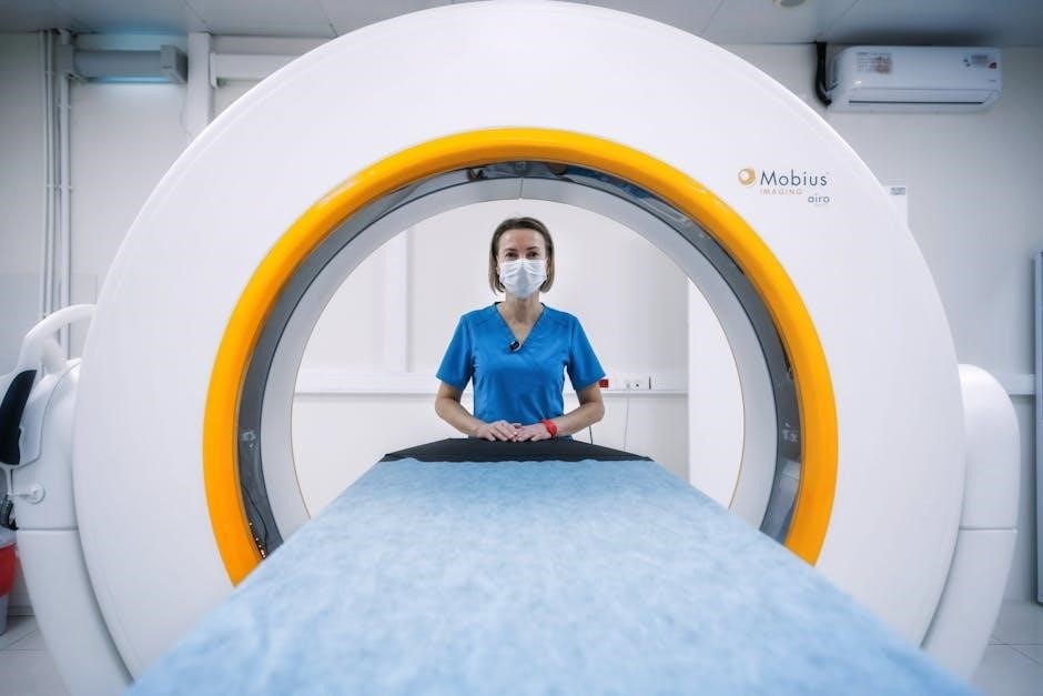
MRI protocols are standardized plans guiding MRI scans, ensuring optimal image quality and diagnostic accuracy. They outline parameters, positioning, and sequences for specific body regions, aiding in consistent, efficient, and safe imaging. Protocols are tailored to clinical needs, ensuring accurate diagnoses and minimizing errors.

Types of MRI Protocols
MRI protocols vary based on body regions and clinical needs. Common types include brain, spine, musculoskeletal, and abdominal scans. Each protocol is tailored to specific anatomical structures, ensuring optimal imaging and diagnostic accuracy. Protocols may also differ by scanner type and manufacturer specifications.
2.1. MSK and Neuro Protocols
MSK (Musculoskeletal) and Neuro MRI protocols are specialized for imaging muscles, bones, and nervous tissues. MSK protocols focus on joints, detecting fractures or soft tissue injuries, using fat-suppressed sequences for clarity. Neuro protocols target brain and spine pathologies, employing high-resolution images to identify abnormalities like tumors or multiple sclerosis. These protocols optimize diagnostic accuracy, ensuring detailed visualization of specific anatomical structures.
Patient Positioning
Patient positioning is critical for clear imaging and diagnostic accuracy. Proper alignment ensures safety, comfort, and reproducibility. Techniques like using positioning blocks and adjusting phase direction optimize image quality, minimizing artifacts. Accurate positioning enhances visualization of anatomical structures, ensuring reliable MRI results.
3.1. Upper and Lower Extremity Positioning
Proper positioning of upper and lower extremities is essential for optimal MRI imaging. For upper extremities, such as the shoulder, elbow, and wrist, patients are typically positioned supine with the affected arm placed in a dedicated coil. The arm should be relaxed, with the elbow slightly flexed to reduce artifact from movement. For the shoulder, the patient’s hand is often placed on the abdomen to maintain alignment. Wrist and finger imaging may require prone or neutral positioning, depending on the clinical indication. Lower extremities, including hips, knees, and ankles, are generally imaged with the patient supine. The legs are positioned straight, with supports used to maintain alignment and reduce movement. For weight-bearing joints like the hips and knees, specific coils and cushions may be used to optimize image quality. Proper alignment ensures the region of interest is centered in the magnetic field, minimizing artifacts and improving diagnostic accuracy. Attention to detail in positioning enhances patient comfort and ensures high-quality MRI results.
Key Parameters
Key MRI parameters include Field of View (FOV), Time to Echo (TE), and Repetition Time (TR). These settings optimize image quality and diagnostic accuracy. FOV defines the imaging area, TE influences contrast, and TR affects scan time and tissue signal characteristics, ensuring proper protocol execution.

4.1. FOV, TE, TR
FOV (Field of View) determines the spatial extent of the MRI image, influencing resolution and scan time. A smaller FOV enhances detail but reduces the area imaged, while a larger FOV captures more anatomy but with lower resolution. TE (Time to Echo) is the time between radiofrequency excitation and signal measurement, affecting tissue contrast. Longer TE emphasizes fluid and soft tissue differences, while shorter TE reduces motion artifacts. TR (Repetition Time) is the interval between successive excitation pulses, impacting scan duration and tissue signal. Shorter TR reduces scan time but may compromise contrast, whereas longer TR improves contrast at the cost of increased scan duration. Balancing these parameters is crucial for optimizing image quality and diagnostic utility in MRI protocols.
Common Indications for MRI Scans
MRI scans are widely used to evaluate a variety of clinical conditions due to their excellent soft-tissue contrast and detailed imaging capabilities. Common indications include musculoskeletal injuries, such as ligament sprains, tendon tears, and cartilage defects. MRI is also frequently used to assess joint disorders, including osteoarthritis, rheumatoid arthritis, and degenerative conditions like spinal disc herniation or stenosis. In neurology, MRI is essential for diagnosing brain injuries, stroke, multiple sclerosis, and structural abnormalities of the central nervous system. For oncologic purposes, MRI helps identify and stage tumors, particularly in soft tissues, liver, and breast. Additionally, it is valuable in evaluating pelvic and abdominal pathologies, such as uterine or prostate conditions. Cardiovascular applications include imaging of vascular structures and heart muscle viability. MRI is also used for preoperative planning in orthopedic and neurosurgical procedures. Its non-invasive nature and lack of ionizing radiation make it a preferred imaging modality for many conditions, especially in pediatric and pregnant patients. Overall, MRI’s versatility and diagnostic precision make it a cornerstone in modern healthcare.

Troubleshooting Common Issues

During MRI protocol planning and execution, various challenges may arise that require prompt resolution. One common issue is motion artifacts, caused by patient movement, which can degrade image quality. To address this, technologists may use faster scanning techniques, patient training, or sedation for anxious individuals. Another issue is claustrophobia, which can be mitigated with open-bore MRI machines or calming strategies like relaxation exercises. Equipment-related problems, such as coil malfunctions or software glitches, may necessitate troubleshooting or equipment replacement. Additionally, artifacts from metal implants or foreign bodies can obscure critical areas, requiring adjustments in pulse sequences or the use of artifact-reducing techniques. Patient positioning errors can also lead to suboptimal imaging, emphasizing the importance of precise alignment and verification before scanning. Furthermore, protocol mismatches, where the wrong sequence is applied, can result in inadequate diagnostic information, highlighting the need for careful planning and verification. Regular quality control checks and technologist training are essential to minimize these issues and ensure high-quality imaging outcomes. By addressing these challenges proactively, MRI protocols can be optimized for both patient comfort and diagnostic accuracy.
Resources for MRI Protocol Planning

Effective MRI protocol planning relies on access to comprehensive resources. Radiologists and technologists often utilize detailed protocol guides provided by manufacturers like Siemens, which offer standardized parameters and sequences for various body regions. Online platforms such as MAGNETOM World provide downloadable protocols, DICOM images, and application tips, including instructional videos. Academic institutions and professional societies, such as the European Society of Radiology, publish guidelines and white papers on specialized imaging techniques. Additionally, textbooks and training manuals offer in-depth explanations of MRI principles and practical examples of protocol implementation. Many healthcare organizations maintain internal databases of updated protocols, ensuring staff access to the latest advancements. Webinars and workshops are another valuable resource, allowing professionals to stay informed about emerging trends and best practices. By leveraging these resources, healthcare providers can ensure that their MRI protocols are evidence-based and optimized for patient care.


Best Practices in MRI Protocol Design
Designing effective MRI protocols involves adherence to best practices that optimize image quality, patient safety, and diagnostic accuracy. Standardization is key, ensuring consistency across scans and reducing variability. Protocols should be tailored to specific clinical indications, balancing parameters like field of view (FOV), repetition time (TR), and echo time (TE) to achieve desired image characteristics. Regular updates are essential to incorporate advancements in technology and clinical knowledge. Collaboration between radiologists, technologists, and physicists ensures comprehensive protocol development. Patient-centered considerations, such as positioning and comfort, should be prioritized to improve compliance and reduce motion artifacts. Additionally, protocols must comply with safety guidelines, including appropriate use of contrast agents and screening for contraindications. Training and education are crucial for staff to understand and implement protocols effectively. Continuous quality improvement through feedback loops and outcome analysis further refines protocol design, ensuring they remain relevant and effective. By following these best practices, healthcare providers can enhance the efficiency and reliability of MRI scans, ultimately improving patient outcomes.
Related Posts

acls exam version c answers pdf
Get ACLS Exam Version C answers in PDF format. Free study guide, practice questions, and instant download. Prepare smarter, not harder!

explaining adhd to a child pdf
Learn how to explain ADHD to children in a simple, engaging way. Download our free PDF guide to help kids understand and manage their ADHD.

john paul jackson books pdf free download
Access John Paul Jackson’s books for free in PDF. Instantly download his spiritual teachings and revelations.